Heart Right and Left Myocardium
Overwhelm conflict
Source: Cranial Nerve Table of GH, 3rd ed., 2025: P108
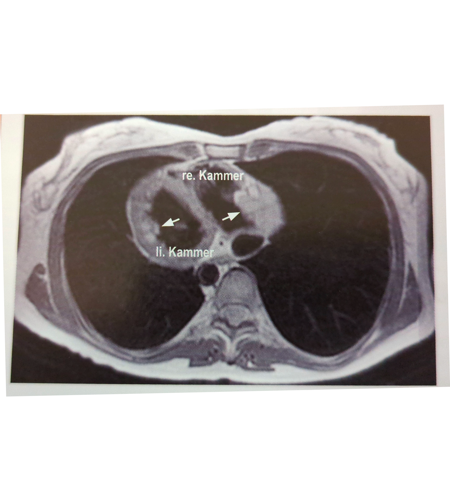
On the CT scan of the heart shown on the left (see below), it can be seen that the outer wall of the right ventricle in this 24-year-old right-handed female patient is markedly thickened. On the inner surface of the outer wall of the left ventricle, a small inward thickening can also be seen (condition following a completed SBS).
The image was taken during surgery whilst the heart was deflated, which is why it is so clearly visible.
The conflict centred primarily on her mother and, to a lesser extent, on her boyfriend.
She wanted to go on holiday to Sicily with her boyfriend.
Her mother was furious and would not allow it.
The conflict had been active for a long time. During the pcl-phase, a muscle sarcoma developed on both sides, accompanied by severe tachycardia.
Unfortunately, the ignorant conventional doctors operated on the right ventricle under the diagnosis of a ‘heart tumour’, ultimately causing the patient’s death.
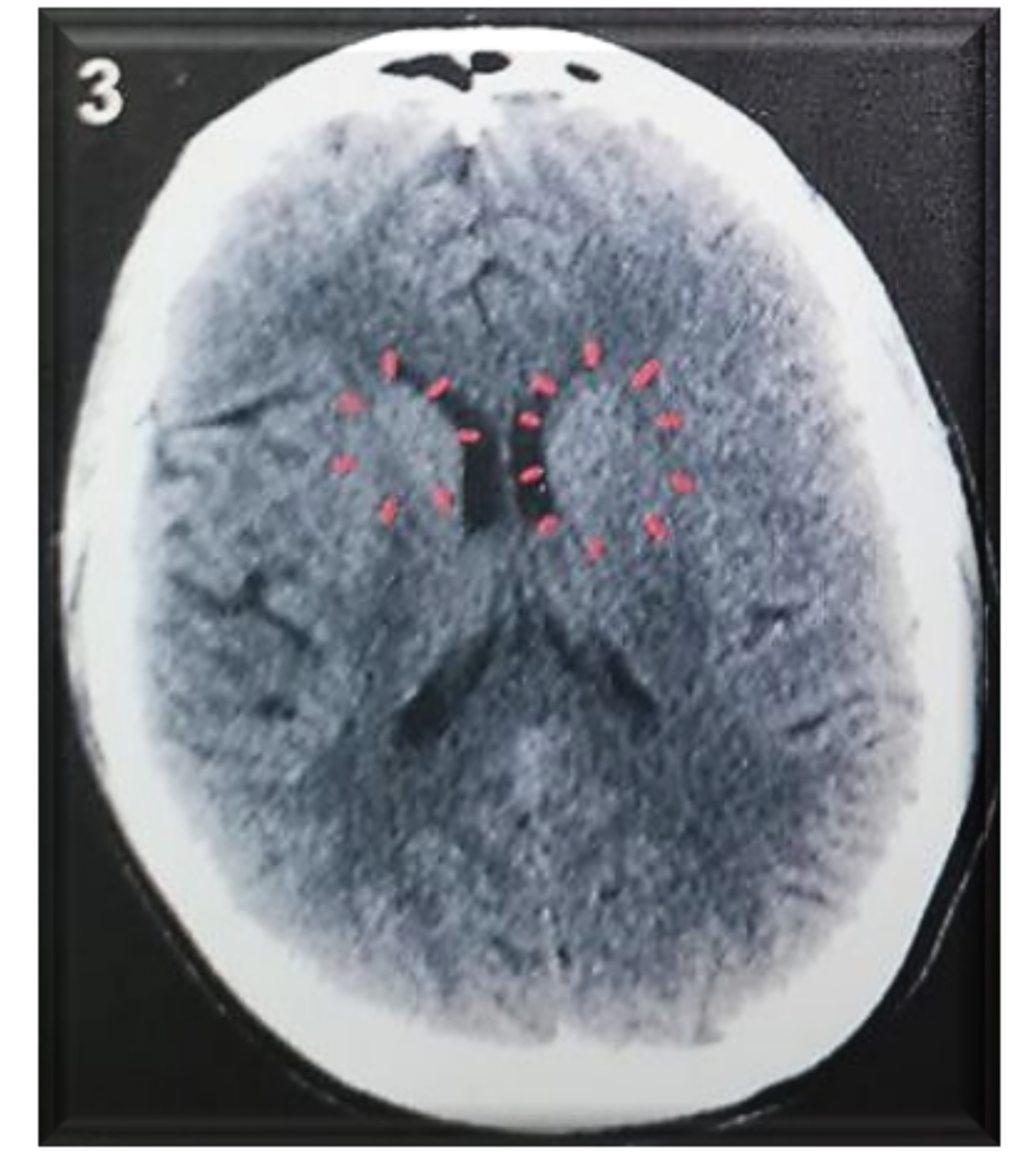
The Brain CT scan shown here reveals the already scarred HHs: on the right, relating to the mother and affecting the right myocardium;
on the left, relating to the boyfriend and affecting the left myocardium.
In both cases, the conflict was twofold: ‘I’ll never manage to soften my mother’s heart’ (right myocardium) and ‘I’ll never manage to go on holiday with my boyfriend without being married’.
We have made the astonishing discovery that many cardiac changes are the result of such muscle-SBS.